human anatomy and physiology laboratory manual 13th edition pdf
Welcome to the study of human anatomy and physiology through this laboratory manual. This resource integrates anatomical structures with physiological functions‚ providing hands-on activities to enhance understanding.
1.1 Overview of the Laboratory Manual
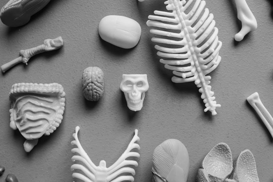
The 13th edition of the Human Anatomy and Physiology Laboratory Manual is a comprehensive guide designed to enhance learning through hands-on exploration. Organized into 13 units‚ it covers key systems of the human body‚ from the skeletal to the integumentary system. Each unit includes detailed exercises‚ dissection guides‚ and interactive activities to reinforce theoretical concepts. The manual emphasizes critical thinking and practical application‚ with visual aids like diagrams and micrographs to aid understanding. It also incorporates real-world scenarios to connect lab work with clinical relevance‚ making it an essential tool for students and instructors alike in anatomy and physiology courses.
1.2 Importance of Lab Work in Anatomy and Physiology
Lab work is essential for understanding the complexities of human anatomy and physiology. It provides hands-on experience‚ allowing students to explore structures and functions firsthand. Through dissection‚ microscopy‚ and experiments‚ learners develop critical thinking and observational skills; Lab activities bridge the gap between theoretical knowledge and practical application‚ enhancing retention and comprehension. They also foster teamwork‚ communication‚ and problem-solving abilities. Additionally‚ lab work prepares students for real-world scenarios in healthcare and research‚ emphasizing the importance of accuracy and safety. By engaging in lab exercises‚ students gain a deeper appreciation for the intricate mechanisms of the human body and its systems.

Laboratory Safety and Best Practices
Laboratory safety is crucial for protecting students and ensuring a productive learning environment. Adhering to safety protocols‚ wearing protective gear‚ and maintaining an organized workspace are essential practices.
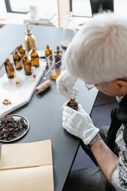
2.1 General Safety Guidelines
Adhering to general safety guidelines is essential in anatomy and physiology labs to prevent accidents and ensure a safe learning environment. Students should wear appropriate personal protective equipment (PPE)‚ such as gloves and lab coats‚ when handling specimens or chemicals. Long hair and loose jewelry should be secured‚ and open-toe shoes avoided. Familiarize yourself with emergency exits‚ fire extinguishers‚ and eyewash stations. Properly label and store chemicals‚ and dispose of biological waste in designated containers. Avoid eating‚ drinking‚ or applying cosmetics in the lab. Follow instructions carefully‚ and never handle sharp instruments carelessly. Report any incidents or spills immediately to the instructor.
2.2 Handling Biological Specimens and Equipment
Proper handling of biological specimens and equipment is critical to maintain safety‚ preserve specimen integrity‚ and ensure accurate laboratory results. Always wear gloves when handling biological materials to prevent exposure to potential pathogens. Use dissecting tools and instruments with care‚ following specific techniques to avoid damage or injury. Biological specimens‚ such as organs or tissues‚ should be stored in appropriate containers and refrigerated when not in use. Equipment like microscopes and scales must be calibrated and cleaned regularly to ensure accuracy and longevity. After use‚ return all materials to their designated storage areas and dispose of waste in labeled biohazard containers.
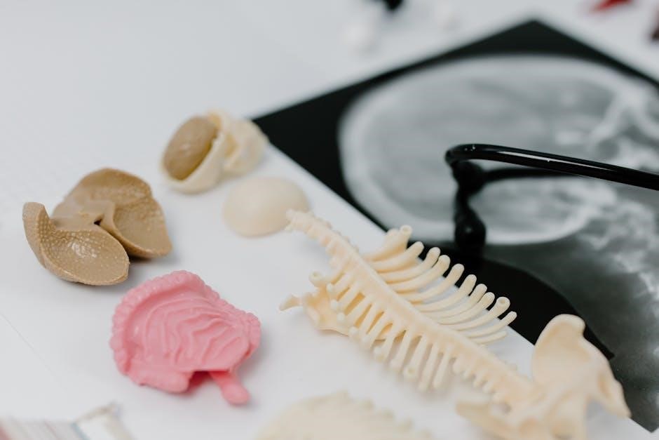
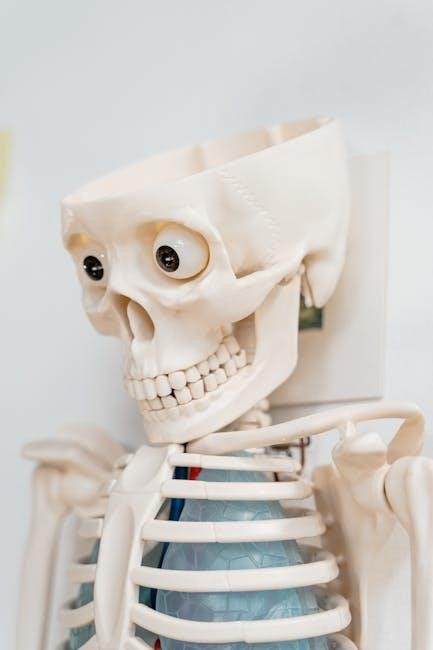
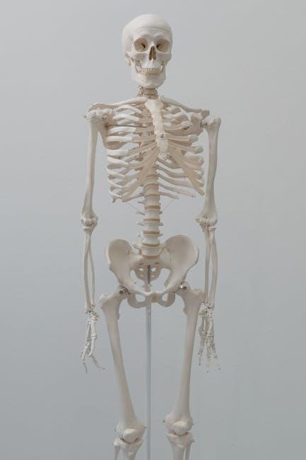
Skeletal System
The skeletal system consists of bones and joints‚ providing structural support‚ protection‚ and facilitating movement. It also produces blood cells and interacts with muscles and nerves.
3.1 Structure and Function of Bones
Bones are rigid‚ calcified tissues forming the skeleton. Their structure includes a compact outer layer and spongy inner tissue. The periosteum covers bones‚ while the cortex and trabeculae provide strength. Bones function in support‚ protection‚ and movement. They also produce blood cells and store minerals like calcium. The medullary cavity contains marrow‚ essential for hematopoiesis. Bones adapt to stress through remodeling‚ ensuring optimal function. This section explores bone histology‚ types‚ and their roles in the body‚ emphasizing practical lab observations to understand their structure and physiological significance.
3.2 Classification and Identification of Joints
Joints‚ or articulations‚ are classified based on their structure and function. Structurally‚ they are fibrous‚ cartilaginous‚ or synovial. Functionally‚ they are synarthroses (immovable)‚ amphiarthroses (slightly movable)‚ or diarthroses (freely movable). Fibrous joints‚ like skull sutures‚ have no joint cavity. Cartilaginous joints‚ such as intervertebral discs‚ allow limited movement. Synovial joints‚ like knees and elbows‚ are the most movable‚ featuring a joint cavity and synovial fluid. In the lab‚ students identify and classify joints using models and diagrams‚ observing structural differences and movement capabilities. This classification aids in understanding joint functions and their roles in human mobility and stability.
Muscular System
The muscular system consists of skeletal‚ smooth‚ and cardiac muscles‚ enabling movement‚ maintaining posture‚ and supporting bodily functions. This section explores their structure‚ functions‚ and interactions in the body.
4.1 Types of Muscles and Their Functions
The human body contains three types of muscles: skeletal‚ smooth‚ and cardiac. Skeletal muscles are voluntary‚ attached to bones‚ and enable movement. Smooth muscles are involuntary‚ found in internal organs‚ and facilitate processes like digestion. Cardiac muscle is specialized for the heart‚ ensuring rhythmic contractions. Each type has distinct structures and functions‚ working together to maintain bodily movements and essential processes. This section explores their unique characteristics‚ roles‚ and interactions within the muscular system.
4.2 Muscle Anatomy and Movement
Muscles are composed of muscle fibers‚ tendons‚ and ligaments‚ working together to facilitate movement. Skeletal muscles attach to bones via tendons‚ enabling voluntary motion. Muscles function by contracting and relaxing‚ with movements occurring at joints. The origin and insertion points of muscles determine their action. Muscles work in pairs as agonists (prime movers) and antagonists (opposing muscles) to produce coordinated movements. Understanding muscle anatomy is crucial for analyzing how the body moves and maintains posture. This section explores the structural components of muscles and their role in movement‚ providing a foundation for laboratory investigations of muscle function and locomotion.
Nervous System
The nervous system consists of the central and peripheral nervous systems‚ coordinating body functions‚ sensory input‚ and motor responses. It enables communication‚ control‚ and adaptation to stimuli through complex neural networks.
5.1 Structure and Function of Neurons
Neurons‚ or nerve cells‚ are specialized for communication and control. They consist of dendrites‚ a cell body‚ and an axon. Dendrites receive signals‚ while the axon transmits them. The cell body contains organelles essential for neuron function. The plasma membrane generates action potentials‚ enabling rapid signal transmission. Neurotransmitters are released at synapses‚ allowing communication with other neurons or target cells. Supporting cells‚ like neuroglial cells‚ assist neuron function. This complex structure allows neurons to integrate and process information‚ enabling sensory perception‚ movement‚ and cognitive processes. Understanding neuron structure and function is fundamental to studying the nervous system’s role in controlling body activities.
5.2 Reflexes and Nerve Impulses
Reflexes are automatic‚ rapid responses to stimuli‚ involving a reflex arc; The reflex arc includes a sensory receptor‚ afferent neuron‚ central nervous system‚ efferent neuron‚ and effector. Nerve impulses‚ or action potentials‚ are electrical and chemical signals that travel along neurons. They are initiated by stimuli‚ propagated by ion exchanges‚ and transmitted to synapses via neurotransmitters. Reflexes ensure immediate responses‚ such as withdrawing a hand from heat‚ without conscious thought. Understanding reflexes and nerve impulses is crucial for grasping how the nervous system maintains homeostasis and enables rapid reactions to environmental changes.

Circulatory System
The circulatory system‚ comprising the heart‚ blood‚ and blood vessels‚ transports oxygen‚ nutrients‚ and hormones while removing waste products‚ essential for maintaining homeostasis and overall health.
6.1 Blood Composition and Blood Types
Blood is a liquid tissue composed of plasma‚ red blood cells (erythrocytes)‚ white blood cells (leukocytes)‚ and platelets. Plasma‚ the liquid portion‚ transports nutrients‚ hormones‚ and waste products. Red blood cells carry oxygen‚ while white blood cells fight infections. Platelets are essential for blood clotting. Blood types are classified based on the presence or absence of specific antigens on red blood cell membranes‚ primarily ABO (A‚ B‚ AB‚ O) and Rh (positive or negative) systems. Understanding blood types is critical for safe transfusions‚ as incompatible types can cause severe reactions. This section explores the components of blood and the significance of blood typing in clinical settings.
6.2 Heart Anatomy and Blood Vessels
The heart is a muscular organ with four chambers: the right and left atria‚ and the right and left ventricles. The septum separates the chambers‚ while valves ensure one-way blood flow. Arteries carry oxygenated blood away from the heart‚ except the pulmonary artery. Veins return deoxygenated blood to the heart‚ except the pulmonary veins. Capillaries enable gas and nutrient exchange. This section examines the heart’s structure‚ including the atrioventricular and semilunar valves‚ and the classification of blood vessels based on their functions and histological features. Understanding these components is essential for grasping circulatory physiology and diagnosing cardiovascular disorders.
Respiratory System
The respiratory system facilitates gas exchange through inhalation and exhalation‚ maintaining oxygen and carbon dioxide balance essential for cellular respiration and overall bodily functions.
7.1 Mechanism of Breathing
Breathing involves the coordinated contraction and relaxation of the diaphragm and intercostal muscles‚ expanding the chest cavity and drawing air into the lungs. Inhalation occurs as the diaphragm descends‚ increasing thoracic volume and lowering air pressure inside the lungs‚ allowing atmospheric air to flow in. Exhalation is passive during relaxed breathing but becomes active during forced expiration‚ with abdominal muscles aiding diaphragm movement. The process is regulated by the autonomic nervous system‚ with the brainstem controlling breathing rate and depth to maintain proper oxygen and carbon dioxide levels in the blood.
The mechanism ensures efficient gas exchange‚ vital for cellular respiration and overall metabolic function.
7.2 Anatomy of the Lungs and Airways
The lungs are cone-shaped organs located in the thoracic cavity‚ protected by the ribcage and diaphragm. Air enters through the trachea‚ which divides into bronchi‚ leading to each lung. Bronchi branch into bronchioles‚ terminating in alveoli‚ where gas exchange occurs. The trachea and bronchi are lined with ciliated epithelium and mucus glands‚ aiding in particle removal. The lungs are divided into lobes‚ with the right lung having three and the left having two. The pleural membrane surrounds each lung‚ reducing friction during breathing. This intricate structure ensures efficient airflow and gas exchange‚ essential for oxygenating the blood and removing carbon dioxide.
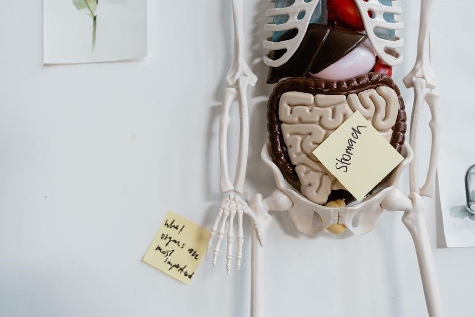
Digestive System
The digestive system processes food into nutrients for absorption. It includes the mouth‚ esophagus‚ stomach‚ intestines‚ and accessory organs like the liver and pancreas‚ enabling digestion and nutrient uptake.
8.1 Structure of the Gastrointestinal Tract
The gastrointestinal (GI) tract is a continuous tube extending from the mouth to the anus. It includes the mouth‚ pharynx‚ esophagus‚ stomach‚ small intestine‚ and large intestine. The mouth ingests and chews food‚ while the pharynx and esophagus transport it to the stomach. The stomach secretes digestive enzymes and acids‚ breaking food into a liquid mixture. The small intestine absorbs most nutrients through villi‚ its finger-like projections. The large intestine absorbs water and electrolytes‚ forming feces for elimination. Each region specializes in specific functions‚ from mechanical digestion to nutrient absorption‚ working together to sustain the body’s nutritional needs.
8.2 Digestive Processes and Enzymes
Digestion involves the breakdown of food into absorbable nutrients. Mechanical digestion begins in the mouth with chewing and continues in the stomach with churning. Chemical digestion relies on enzymes that break down specific nutrients. Amylase in saliva and pancreatic juice targets carbohydrates‚ while lipase in the stomach and small intestine handles fats. Proteases like pepsin and trypsin digest proteins into amino acids. These enzymes function optimally at specific pH levels‚ ensuring efficient nutrient absorption in the small intestine. Understanding these processes and enzymes is crucial for appreciating how the body maintains proper nutrition and overall health.
Urinary System
The urinary system filters blood‚ removes waste‚ and regulates electrolytes and fluid balance. It includes kidneys‚ ureters‚ bladder‚ and urethra‚ essential for maintaining homeostasis and overall health.
9.1 Kidney Structure and Function
The kidneys are bean-shaped organs located in the lower back‚ responsible for filtering blood to remove waste and excess substances. Each kidney contains over a million nephrons‚ the functional units. Nephrons consist of a glomerulus‚ a cluster of capillaries‚ and a renal tubule. Blood enters the glomerulus‚ where filtration occurs‚ and the tubule reabsorbs or secretes substances to regulate electrolytes‚ pH‚ and fluid balance. The kidneys also produce hormones like erythropoietin and renin‚ essential for red blood cell production and blood pressure regulation. Proper kidney function is vital for maintaining homeostasis and overall health‚ making them a critical component of the urinary system.
9.2 Urine Formation and Excretion
Urine formation begins with filtration in the glomerulus‚ where blood is filtered to produce filtrate. The renal tubules then reabsorb water‚ nutrients‚ and ions‚ while secreting waste products. This process regulates blood composition and maintains homeostasis. Urine is transported to the bladder via the ureters for storage. The bladder expands to hold urine until excretion occurs through the urethra. Nervous system control‚ including the autonomic nervous system‚ regulates this process. Proper urine formation and excretion are essential for removing waste and excess substances‚ ensuring overall health and maintaining fluid balance in the body.
Endocrine System
The endocrine system is a network of glands producing hormones that regulate metabolism‚ growth‚ and reproductive processes‚ essential for maintaining homeostasis and overall bodily functions.
10.1 Major Endocrine Glands and Hormones
The endocrine system comprises several key glands that produce hormones regulating various bodily functions. The pituitary gland‚ often called the “master gland‚” controls other endocrine glands and regulates growth and metabolism. The thyroid gland produces thyroxine‚ essential for metabolism and energy levels. The adrenal glands release adrenaline and cortisol‚ influencing stress responses and blood sugar regulation. The pancreas secretes insulin and glucagon to manage blood glucose levels. The ovaries produce estrogen and progesterone‚ crucial for reproductive processes‚ while the testes produce testosterone‚ regulating male reproductive and sexual functions. These glands and their hormones are vital for maintaining homeostasis and overall health.
10.2 Regulation of Hormonal Balance
Hormonal balance is maintained through complex feedback mechanisms involving the endocrine system‚ nervous system‚ and target organs. The hypothalamus monitors hormone levels and triggers the pituitary gland to release or inhibit hormones‚ ensuring equilibrium. Negative feedback loops‚ such as those involving insulin and cortisol‚ prevent excessive hormone production. Positive feedback loops‚ like oxytocin during childbirth‚ amplify responses when needed. The autonomic nervous system also influences hormone secretion‚ while factors like stress‚ nutrition‚ and sleep impact balance. Proper regulation is crucial for metabolic‚ reproductive‚ and stress responses‚ ensuring overall physiological harmony and health.
Reproductive System
The reproductive system encompasses male and female structures essential for producing sex cells and supporting development. This laboratory manual provides hands-on exploration of anatomical and physiological processes.
11.1 Male Reproductive Anatomy
The male reproductive system consists of the testes‚ epididymis‚ vas deferens‚ seminal vesicles‚ prostate gland‚ and penis. The testes produce sperm and testosterone‚ while the epididymis stores and matures sperm. The vas deferens transports sperm during ejaculation. Seminal vesicles and the prostate gland secrete fluids that make up semen. The penis delivers sperm during ejaculation. This laboratory manual provides detailed dissections and histological examinations to explore these structures‚ emphasizing their roles in sperm production and delivery. Hands-on activities include identifying external and internal reproductive organs‚ analyzing sperm samples‚ and understanding the functional relationships between these components.
11.2 Female Reproductive Anatomy
The female reproductive system includes the ovaries‚ fallopian tubes‚ uterus‚ cervix‚ and vagina. The ovaries produce eggs and hormones like estrogen and progesterone. The fallopian tubes facilitate fertilization‚ while the uterus supports embryonic development during pregnancy. The cervix connects the uterus to the vagina‚ which serves as the birth canal and pathway for menstrual flow. This laboratory manual provides detailed explorations of female reproductive structures through dissection and histological studies. Activities include identifying external and internal reproductive organs‚ examining the menstrual cycle’s physiological changes‚ and understanding the functional relationships between these components. Hands-on exercises enhance comprehension of female reproductive health and anatomy.
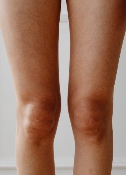
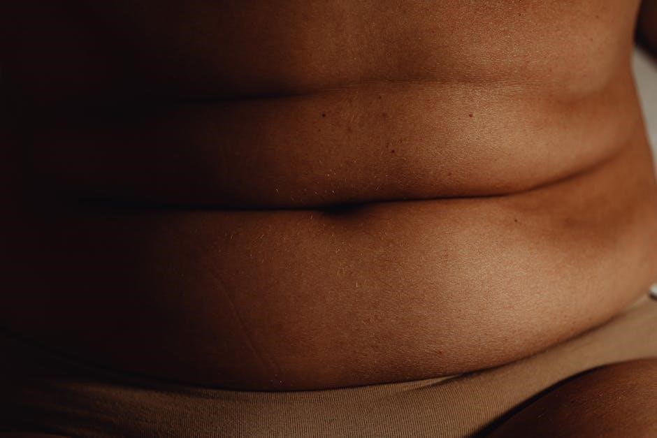
Integumentary System
The integumentary system‚ comprising skin and its accessories‚ protects‚ regulates‚ and senses stimuli. This section explores skin structure‚ function‚ and laboratory investigations‚ enhancing understanding of its vital roles.
12.1 Skin Structure and Function
The skin is the body’s largest organ‚ consisting of the epidermis‚ dermis‚ and hypodermis. The epidermis‚ outermost layer‚ protects against external factors and contains keratinocytes and melanocytes. The dermis‚ underlying layer‚ contains blood vessels‚ nerve endings‚ and glands‚ regulating body temperature and sensation. The hypodermis‚ deepest layer‚ anchors the skin and stores fat. Skin functions include protection‚ thermoregulation‚ sensation‚ and aiding in vitamin D production. Laboratory exercises explore skin layers‚ appendages‚ and sensory receptors‚ providing hands-on understanding of its complex roles in maintaining homeostasis and overall health.
12.2 Accessories of the Skin
The skin’s accessories include hair‚ nails‚ sweat glands‚ and sebaceous glands. Hair provides protection and aids in sensation‚ while nails protect the tips of fingers and toes. Sweat glands regulate body temperature through perspiration‚ and sebaceous glands produce sebum to moisturize the skin. These structures contribute to thermoregulation‚ protection‚ and overall bodily functions. Laboratory exercises often involve identifying these accessories and understanding their roles in maintaining skin health and function. Practical activities may include observing hair and nail samples under a microscope or analyzing sweat and sebaceous gland secretions to appreciate their importance in human anatomy and physiology.
Mastering human anatomy and physiology requires the integration of theoretical knowledge with practical lab experiences‚ equipping students with essential skills applicable in real-world healthcare scenarios.
13.1 Summary of Key Concepts
This laboratory manual provides a comprehensive exploration of human anatomy and physiology‚ emphasizing the interconnection between structure and function. Through hands-on activities‚ students gain practical skills in identifying anatomical structures‚ understanding physiological processes‚ and applying scientific methods. Key concepts include the skeletal‚ muscular‚ nervous‚ circulatory‚ respiratory‚ digestive‚ urinary‚ endocrine‚ and reproductive systems‚ as well as the integumentary system. The manual also highlights the importance of laboratory safety‚ proper use of equipment‚ and ethical handling of biological specimens. By integrating theoretical knowledge with experimental learning‚ this resource fosters a deeper understanding of human biology and its applications in healthcare and research.
13.2 Application of Lab Skills in Real-World Scenarios
The skills acquired through this laboratory manual are invaluable in real-world applications‚ particularly in healthcare‚ research‚ and education. Healthcare professionals rely on anatomical knowledge to diagnose and treat conditions‚ while researchers use physiological insights to develop new treatments. Educators apply these skills to teach future generations of scientists and medical practitioners. Additionally‚ fitness and wellness professionals benefit from understanding muscle function and movement to design safe exercise programs. The hands-on experience gained from this manual prepares students to confidently transition into these roles‚ ensuring they can apply their knowledge effectively in diverse professional settings.
Related Posts
pathfinder 2e remaster pdf
Dive into the epic world of Pathfinder 2nd Edition! Download the complete remaster PDF and unleash thrilling quests, powerful characters, & endless possibilities. Get yours today!

romans study guide pdf
Struggling with Roman history? Get our FREE, easy-to-use study guide in PDF format! Conquer emperors, battles, and culture – download now & boost your grade!

iris kelly doesn’t date pdf
Explore the captivating writings of Iris Kelly! Get instant access to PDFs of her work and discover thought-provoking content. Download now and immerse yourself in her ideas. **Iris Kelly** awaits!