neonatal ventilator settings pdf
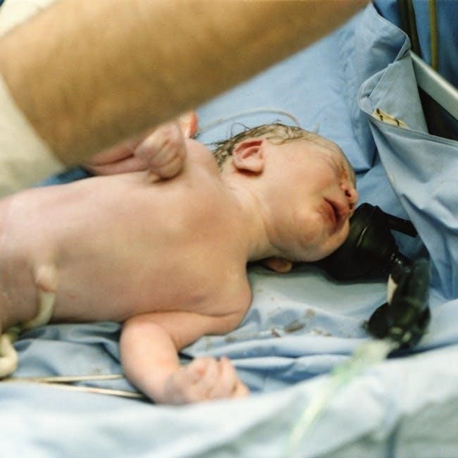
Neonatal ventilator settings are critical for supporting respiratory function in newborns with respiratory distress. Various modes‚ such as HFOV and synchronized ventilation‚ are used to optimize gas exchange. Key parameters like PEEP‚ tidal volume‚ and inspiratory pressure must be precisely set to ensure effective ventilation while minimizing complications. Proper setup and monitoring are essential for improving outcomes.
- Modes of ventilation: HFOV‚ synchronized ventilation.
- Parameters: PEEP‚ tidal volume‚ inspiratory pressure.
- Importance: Ensures proper gas exchange and minimizes complications.
The goal is to provide tailored respiratory support‚ adapting to the neonate’s specific needs for optimal care.

Key Concepts and Terminology in Neonatal Ventilation
Understanding key terms is vital for effective neonatal ventilation. Modes like HFOV and NIV‚ parameters such as PEEP and tidal volume‚ and terms like LBW and NICU are essential. These concepts ensure personalized respiratory support‚ adapting to each neonate’s needs for optimal care and outcomes.
- Modes: HFOV‚ NIV‚ synchronized ventilation.
- Parameters: PEEP‚ tidal volume‚ inspiratory pressure.
- Abbreviations: LBW (Low Birth Weight)‚ NICU (Neonatal Intensive Care Unit).
2.1. Modes of Neonatal Ventilation
Neonatal ventilation involves various modes tailored to the infant’s respiratory needs. Common modes include High Frequency Oscillatory Ventilation (HFOV)‚ which delivers rapid‚ small breaths to maintain lung volume‚ and Non-Invasive Ventilation (NIV)‚ such as CPAP‚ which supports breathing without intubation; Synchronized ventilation modes‚ like SIMV‚ align with the infant’s spontaneous breaths‚ reducing discomfort and improving effectiveness. Each mode is selected based on the neonate’s condition‚ such as respiratory distress syndrome or meconium aspiration.
- HFOV: Ideal for severe lung conditions‚ as it minimizes lung injury.
- NIV: Prevents intubation and is used for mild respiratory support.
- SIMV: Enhances patient-ventilator synchrony‚ promoting comfort and effectiveness.
Understanding these modes ensures personalized respiratory support‚ optimizing outcomes for critically ill neonates.
2.2. Importance of Ventilator Settings in Neonatal Care
Accurate ventilator settings are crucial in neonatal care to support respiratory function and prevent complications. Proper settings ensure effective gas exchange‚ maintaining adequate oxygenation and carbon dioxide removal. Incorrect parameters can lead to lung injury or inadequate ventilation‚ emphasizing the need for precise adjustments based on patient response and clinical guidelines.
- Effective Gas Exchange: Maintains proper oxygen and carbon dioxide levels.
- Prevention of Complications: Avoids lung injury and ensures optimal ventilation.
- Customization: Settings are tailored to the neonate’s specific respiratory needs.
Regular monitoring and timely adjustments are essential to ensure the ventilator supports the neonate’s recovery effectively‚ minimizing risks and improving outcomes.
2.3. Common Ventilation Parameters and Their Significance
In neonatal ventilation‚ several key parameters are crucial for ensuring effective respiratory support. These settings are tailored to the neonate’s specific needs and are continuously monitored to optimize outcomes. The primary parameters include:
- Positive End-Expiratory Pressure (PEEP): Maintains lung inflation at the end of exhalation‚ preventing alveolar collapse and improving oxygenation.
- Peak Inspiratory Pressure (PIP): The highest pressure applied during inhalation‚ ensuring adequate tidal volume delivery while minimizing lung injury risk.
- Tidal Volume (VT): The volume of air delivered with each breath‚ critical for maintaining effective gas exchange without causing overdistension;
- Respiratory Rate: The number of breaths per minute‚ synchronized with the neonate’s breathing efforts to enhance comfort and efficiency.
- Inspiratory Time: The duration of the inspiratory phase‚ ensuring sufficient time for lung inflation while preventing prolonged pressures.
Understanding and adjusting these parameters is vital for optimizing ventilation support‚ preventing complications‚ and promoting lung health in neonates. Continuous monitoring and tailored adjustments ensure the best possible outcomes for critically ill newborns.

Physiological Principles of Neonatal Ventilation
Neonatal ventilation relies on understanding lung mechanics‚ gas exchange‚ and neural breathing control. Neonates have delicate lungs with high compliance and low recoil. Surfactant deficiency and immature respiratory muscles require careful ventilator settings to maintain adequate oxygenation and carbon dioxide removal‚ ensuring proper physiological support for their developing respiratory system.
- Lung mechanics: High compliance‚ low recoil.
- Gas exchange: Maintaining oxygenation and CO2 removal.
- Neural control: Immature respiratory centers influence breathing patterns.
3.1. Lung Mechanics in Neonates
Understanding lung mechanics is crucial for effective neonatal ventilation. Neonates have highly compliant lungs but low recoil‚ making them prone to atelectasis. Surfactant deficiency‚ common in preterm infants‚ increases surface tension‚ leading to difficulty in lung expansion. This necessitates careful ventilator settings to maintain adequate lung volumes and prevent injury.
The small size of neonatal airways results in higher resistance‚ requiring precise pressure and flow adjustments. Tidal volumes must be tailored to avoid volutrauma‚ while ensuring sufficient gas exchange. Ventilator parameters like PEEP and inspiratory pressure are critical to counteract the effects of surfactant deficiency and maintain functional residual capacity.
- Compliance and Recoil: High compliance but low elastic recoil increases susceptibility to lung collapse.
- Surfactant Deficiency: Leads to increased surface tension‚ complicating lung expansion and gas exchange.
- Airway Resistance: Narrow airways increase resistance‚ affecting ventilation efficiency.
- Ventilator Adjustments: PEEP and inspiratory pressure settings are vital to maintain lung volumes and prevent injury.
Optimizing lung mechanics through appropriate ventilator settings is essential to support the neonate’s respiratory function and minimize the risk of long-term pulmonary complications.
3.2. Gas Exchange and Oxygenation in Neonatal Ventilation
Gas exchange and oxygenation are fundamental goals of neonatal ventilation. Effective ventilation ensures adequate oxygenation and carbon dioxide removal‚ critical for tissue perfusion and metabolic function. Neonates‚ especially preterm infants‚ often struggle with impaired gas exchange due to surfactant deficiency‚ lung immaturity‚ and reduced alveolar surface area.
Tidal volume and positive end-expiratory pressure (PEEP) are key parameters influencing gas exchange. Proper adjustment of these settings ensures optimal alveolar recruitment while minimizing the risk of volutrauma or atelectasis. Oxygen saturation (SpO2) and partial pressure of carbon dioxide (PaCO2) are closely monitored to assess ventilatory efficacy and guide adjustments.
- Tidal Volume: Affects alveolar ventilation and gas exchange efficiency.
- PEEP: Maintains alveolar recruitment‚ preventing collapse and improving oxygenation.
- Oxygen Saturation (SpO2): Serves as a non-invasive marker of oxygenation status.
- PaCO2 Levels: Reflect ventilatory adequacy and respiratory function.
Optimizing gas exchange requires balancing ventilator settings to meet the neonate’s changing physiological needs‚ ensuring adequate oxygen delivery while avoiding hyperoxia or hypoventilation. Continuous monitoring and tailored adjustments are essential to support the neonate’s respiratory and overall clinical outcomes.
3.3. Neural Control of Breathing in Neonates
Neural control of breathing in neonates is a complex process that is still maturing. The respiratory center‚ located in the brainstem‚ regulates breathing patterns through signals to the diaphragm and accessory muscles. In neonates‚ especially preterm infants‚ this system is highly sensitive and can be influenced by various factors such as sleep-wake cycles‚ environmental stimuli‚ and clinical conditions like hypoxia or hypercapnia.
Neonates exhibit irregular breathing patterns‚ characterized by periods of rapid breathing interspersed with brief pauses. This immaturity can lead to respiratory instability‚ often necessitating ventilatory support. The development of neural control mechanisms is crucial for transitioning from mechanical ventilation to independent breathing. Understanding these neural processes aids in optimizing ventilator settings to synchronize with the neonate’s intrinsic respiratory efforts.
- Respiratory Center: Located in the brainstem‚ it regulates breathing rhythm and depth.
- Neonatal Breathing Patterns: Often irregular‚ with fluctuations in rate and depth.
- Maturation Process: Essential for achieving stable‚ independent respiration.
Assessing the neonate’s neural respiratory control is vital for determining the appropriate level of ventilatory support and weaning strategies. Enhancing neural adaptability and stability through synchronized ventilation can significantly improve respiratory outcomes and reduce long-term complications.

Clinical Assessment and Monitoring in Neonatal Ventilation
Clinical assessment and monitoring are crucial in neonatal ventilation to ensure effective respiratory support. Regular evaluation of vital signs‚ chest movement‚ and breath sounds helps identify ventilation adequacy. Advanced tools like pulse oximetry and capnography monitor oxygenation and carbon dioxide levels‚ guiding adjustments in ventilator settings for optimal patient outcomes.
- Vital Signs: Heart rate‚ respiratory rate‚ and oxygen saturation;
- Physical Assessment: Chest movement‚ breath sounds‚ and work of breathing.
- Advanced Monitoring: Pulse oximetry‚ capnography‚ and blood gas analysis.
Continuous monitoring ensures timely interventions‚ improving ventilation efficacy and reducing complications in critically ill neonates.
4.1. Patient Assessment for Ventilator Support
Patient assessment for ventilator support is a critical step in neonatal care‚ ensuring that respiratory intervention is both necessary and appropriately tailored. This process involves evaluating the neonate’s respiratory status‚ including signs of distress such as tachypnea‚ retractions‚ and grunting. Clinicians assess oxygenation and ventilation through pulse oximetry‚ capnography‚ and blood gas analysis to determine the severity of respiratory compromise.
The neonate’s overall clinical condition‚ including gestational age‚ birth weight‚ and any underlying conditions‚ is considered. Vital signs‚ such as heart rate and respiratory rate‚ are monitored to identify deviations from normal ranges. Physical examination focuses on chest movement‚ breath sounds‚ and the presence of nasal flaring‚ which indicate the effort and effectiveness of breathing.
- Respiratory Signs: Tachypnea‚ retractions‚ grunting‚ and nasal flaring.
- Oxygenation and Ventilation: Pulse oximetry‚ capnography‚ and blood gas analysis.
- Overall Condition: Gestational age‚ birth weight‚ and underlying medical conditions.
Advanced monitoring tools‚ such as esophageal manometry‚ may be used to assess lung mechanics and guide ventilator settings. The goal of this comprehensive assessment is to identify neonates who would benefit from ventilatory support and to tailor the intervention to their specific needs‚ ensuring optimal respiratory outcomes. Early and accurate assessment is key to preventing complications and improving the quality of care for critically ill neonates.
4.2. Monitoring Techniques and Tools in Neonatal Ventilation
Monitoring techniques and tools are essential in neonatal ventilation to ensure effective and safe respiratory support. Continuous assessment of the neonate’s respiratory status helps in making timely adjustments to ventilator settings. Pulse oximetry is widely used to monitor oxygen saturation (SpO2) and ensure adequate oxygenation‚ while capnography measures end-tidal CO2 levels‚ providing insights into ventilation effectiveness.
Blood gas analysis is a cornerstone in evaluating gas exchange‚ helping to assess the adequacy of oxygenation and ventilation. This test measures pH‚ PaCO2‚ and PaO2 levels‚ guiding adjustments in ventilator parameters such as tidal volume and respiratory rate. Additionally‚ chest radiographs are used to confirm endotracheal tube placement and assess lung expansion and aeration.
- Pulse Oximetry: Monitors SpO2 to ensure proper oxygenation.
- Capnography: Measures end-tidal CO2 to assess ventilation.
- Blood Gas Analysis: Evaluates pH‚ PaCO2‚ and PaO2 for gas exchange.
- Chest Radiographs: Confirms tube placement and lung status.
Advanced tools like esophageal manometry and respiratory inductance plethysmography provide detailed information on lung mechanics and breathing patterns‚ aiding in the optimization of ventilator settings. Continuous monitoring ensures that the neonate receives the most appropriate level of respiratory support‚ minimizing the risk of complications and promoting better outcomes. Regular review of these monitoring tools by healthcare providers is crucial for maintaining effective and safe ventilation therapy in neonates.

Setting Up the Neonatal Ventilator
Setting up the neonatal ventilator involves selecting appropriate modes and inputting parameters based on the neonate’s condition. Modes like HFOV or synchronized ventilation are chosen‚ and settings such as PIP‚ PEEP‚ rate‚ and tidal volume are entered. Proper setup ensures effective respiratory support and minimizes complications. Always confirm settings align with clinical guidelines and the neonate’s needs.
- Selecting ventilation modes (e.g.‚ HFOV‚ synchronized).
- Setting parameters: PIP‚ PEEP‚ rate‚ tidal volume.
- Ensuring settings match clinical guidelines and patient needs.
Customization is key to providing tailored respiratory support and optimizing outcomes for the neonate.
5.1. Initial Ventilator Settings for Neonates
Initial ventilator settings for neonates are tailored to their specific respiratory needs‚ ensuring adequate gas exchange while minimizing lung injury. Typically‚ a pressure-limited ventilator is used‚ starting with a peak inspiratory pressure (PIP) of 20-25 cm H2O and a positive end-expiratory pressure (PEEP) of 4-6 cm H2O. The respiratory rate is often set between 40-60 breaths per minute‚ with an inspiratory time of 0.3-0.5 seconds. Tidal volumes should range from 4-6 mL/kg to prevent volutrauma.
The fraction of inspired oxygen (FiO2) is initially set to 1.0 but adjusted based on oxygen saturation targets (usually 88-96%). For neonates with respiratory distress syndrome (RDS)‚ surfactant therapy may be indicated to improve lung compliance. In cases of severe respiratory failure‚ high-frequency oscillatory ventilation (HFOV) may be considered‚ with mean airway pressures (MAP) set to maintain lung volume.
- PIP: 20-25 cm H2O
- PEEP: 4-6 cm H2O
- Rate: 40-60 breaths per minute
- Inspiratory time: 0.3-0.5 seconds
- Tidal volume: 4-6 mL/kg
- FiO2: 1.0 initially‚ adjusted as needed
These settings are guidelines and must be individualized based on the neonate’s response and underlying condition. Continuous monitoring of vital signs‚ blood gases‚ and clinical improvement is essential to guide adjustments. Always consult clinical guidelines and expert recommendations for specific cases.
5.2. Adjusting Ventilator Parameters Based on Patient Response
Adjusting ventilator parameters in neonates requires careful monitoring of the patient’s response to current settings. Key indicators for adjustment include arterial blood gas (ABG) results‚ oxygen saturation (SpO2)‚ and clinical signs such as respiratory effort and chest movement. If the neonate remains hypoxemic (low SpO2)‚ increasing the fraction of inspired oxygen (FiO2) or positive end-expiratory pressure (PEEP) may be necessary. Conversely‚ if oxygen levels are too high‚ gradual reduction of FiO2 and PEEP should be considered to prevent oxidative stress.
Carbon dioxide levels (PaCO2) from ABGs guide adjustments in ventilation rate or tidal volume. Elevated PaCO2 indicates insufficient ventilation‚ necessitating an increase in respiratory rate or tidal volume. Low PaCO2 may suggest over-ventilation‚ leading to reduced settings to prevent alkalosis. Additionally‚ clinical signs like retractions or grunting signal the need for increased respiratory support‚ possibly through higher PIP or transitioning to alternative modes like HFOV.
- Monitor ABGs‚ SpO2‚ and clinical signs.
- Adjust FiO2 and PEEP for oxygenation.
- Tweak rate or tidal volume based on PaCO2 levels.
- Watch for signs of respiratory distress;
These adjustments must be made cautiously‚ with incremental changes‚ to avoid sudden shifts in the neonate’s respiratory status. Continuous assessment and documentation are crucial to ensure optimal ventilatory support and minimize complications. Always refer to institutional protocols and consult with neonatal specialists when uncertain.

Common Complications and Management in Neonatal Ventilation
Common complications in neonatal ventilation include bronchopulmonary dysplasia (BPD)‚ ventilator-induced lung injury (VILI)‚ and air leaks. Management involves optimizing ventilation parameters‚ using non-invasive methods when possible‚ and ensuring minimal oxygen exposure. Regular monitoring and early intervention are crucial to mitigate long-term respiratory and developmental issues in neonates.
- BPD: Chronic lung disease from prolonged ventilation.
- VILI: Lung damage due to excessive pressure or volume.
- Air leaks: Pneumothorax or pulmonary interstitial emphysema.
Strategies focus on lung-protective ventilation and individualized care to improve outcomes.
6.1. Complications Associated with Neonatal Ventilation
Neonatal ventilation‚ while life-saving‚ carries risks of complications that can significantly impact a newborn’s health. Common issues include bronchopulmonary dysplasia (BPD)‚ a chronic lung disease resulting from prolonged mechanical ventilation and oxygen exposure. Ventilator-induced lung injury (VILI) can occur due to excessive pressure or tidal volumes‚ leading to inflammation and tissue damage. Air leaks‚ such as pneumothorax or pulmonary interstitial emphysema‚ are additional concerns and may require immediate intervention.
Other complications include respiratory acidosis‚ hypoxemia‚ and tracheal trauma from endotracheal tubes. Long-term respiratory and neurodevelopmental delays are potential sequelae‚ particularly in preterm infants. These complications underscore the need for precise ventilator settings and continuous monitoring to minimize adverse outcomes.
- BPD: Chronic lung disease linked to prolonged ventilation;
- VILI: Lung damage from mechanical stress during ventilation.
- Air leaks: Pneumothorax or pulmonary interstitial emphysema.
- Respiratory acidosis: Elevated CO2 levels due to inadequate ventilation.
- Tracheal trauma: Injury from endotracheal intubation.
Understanding these complications is critical for optimizing ventilator care and improving neonatal outcomes.
Related Posts

logic puzzles pdf with answers
Sharpen your mind with our collection of free, downloadable logic puzzles in PDF format! Perfect for all ages – test your skills & find the answers. Download now!

13 elliott wave patterns pdf
Unlock the secrets of the market! Download our comprehensive PDF guide to 13 Elliott Wave patterns & start predicting price movements with confidence. Learn now!

6th grade iready math book pdf
Need a 6th grade iReady Math book PDF? Get instant access to the complete curriculum! Boost your grades & conquer math with our easy-to-download resource. iReady made simple!